

01
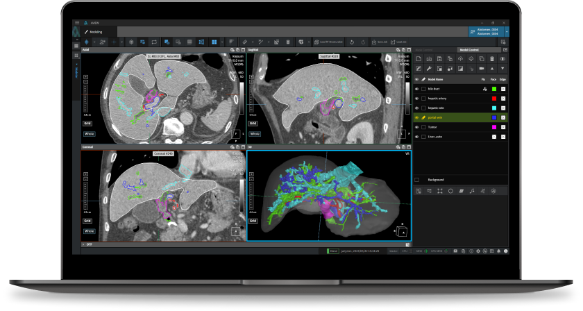
Key features
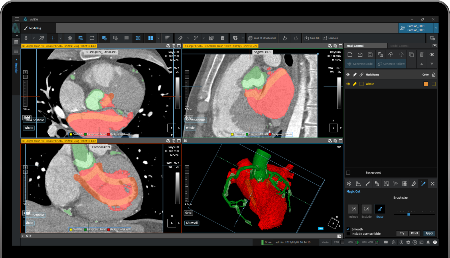
Brush
Add or remove free drawing dots or lines from masks
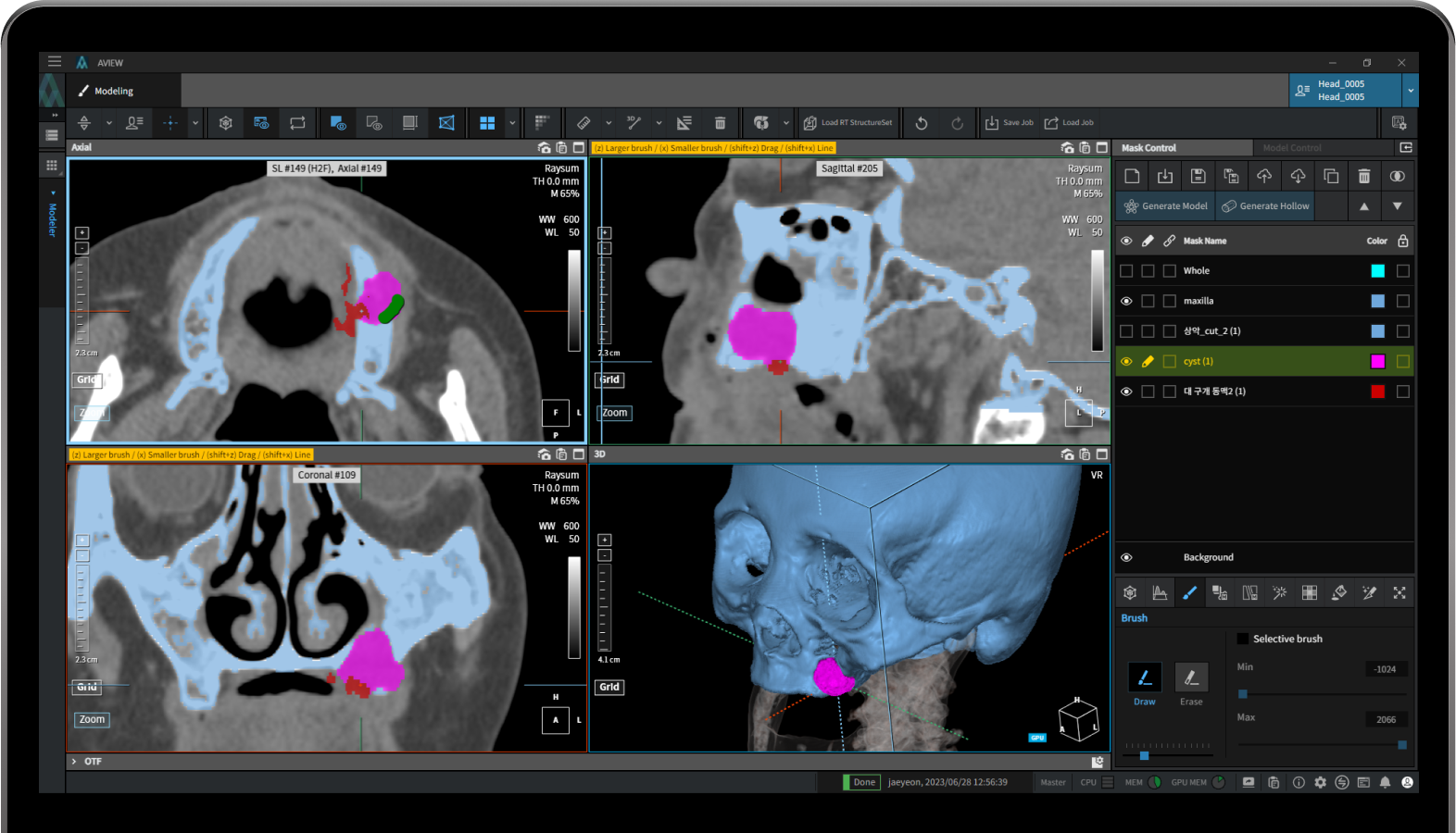
Pick
Click to separate linked masks.
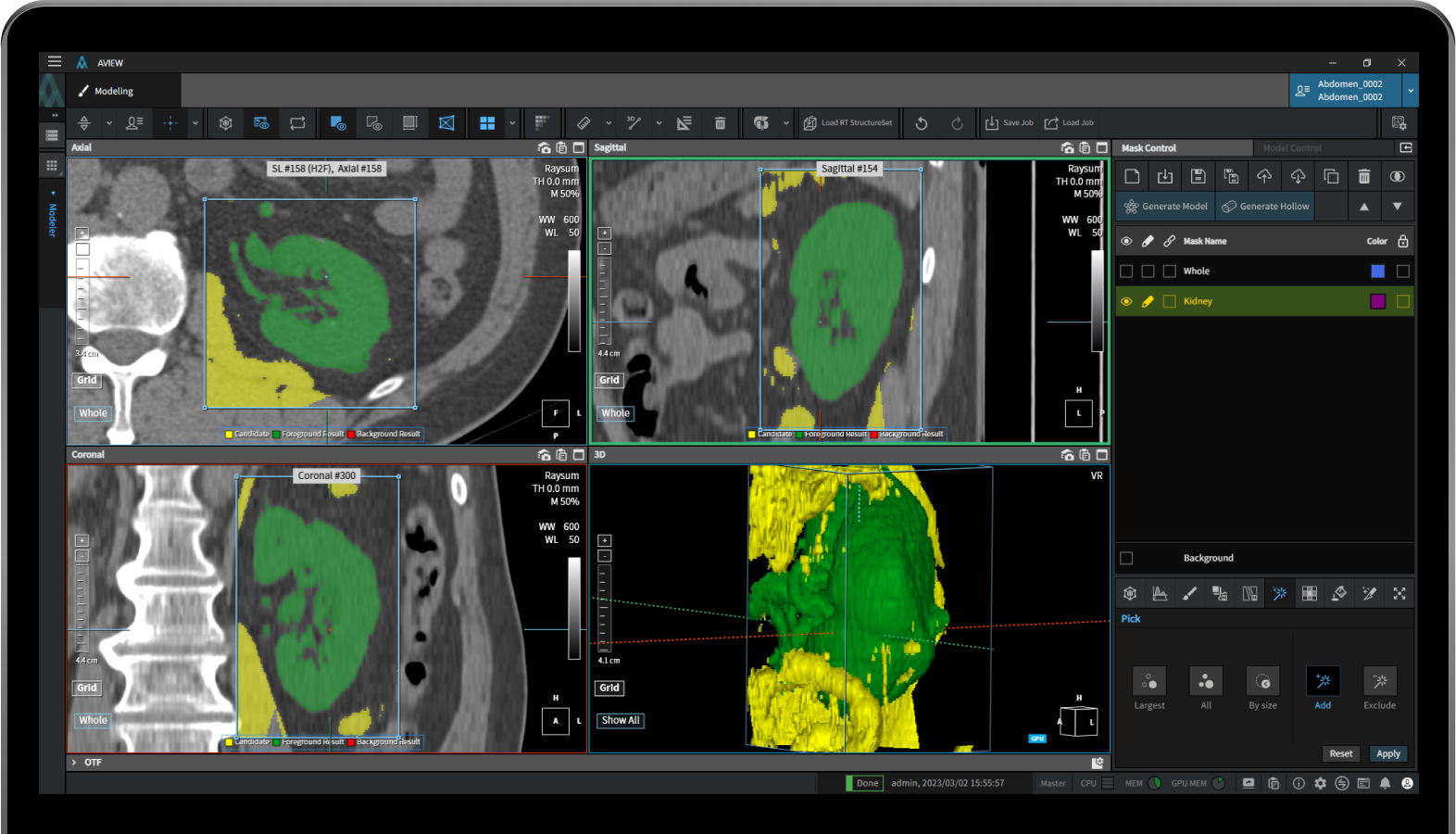
Sculpt
Adjust straight, curved, and free-draw lines for desired body parts from masks.
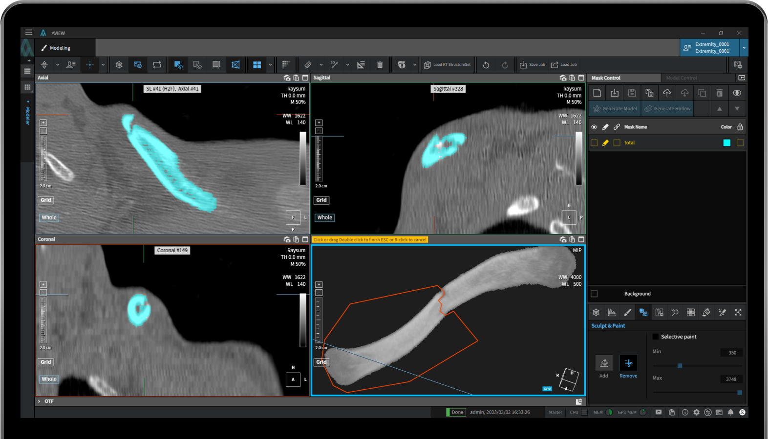
Magic cut segmentation
Automatically segment boundaries of body parts by drawing reference lines on a slice.
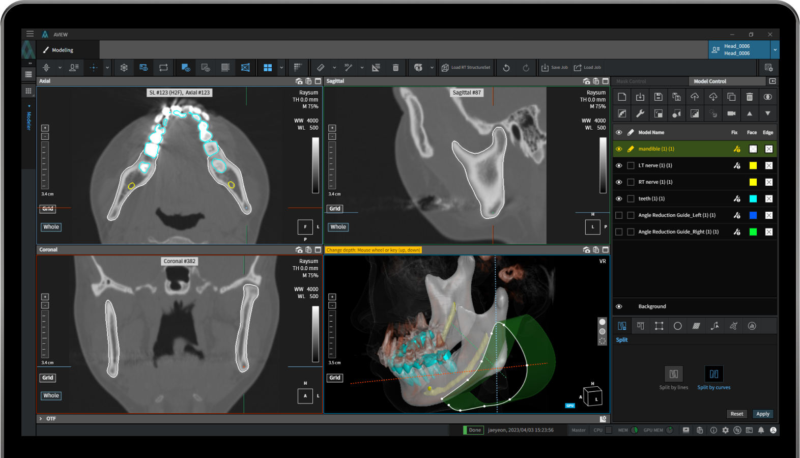
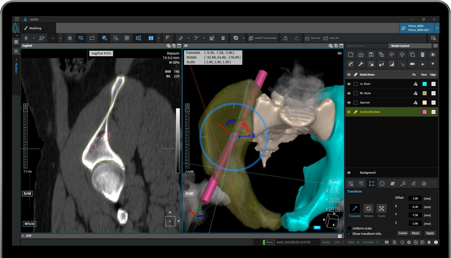
Split Model
Split areas
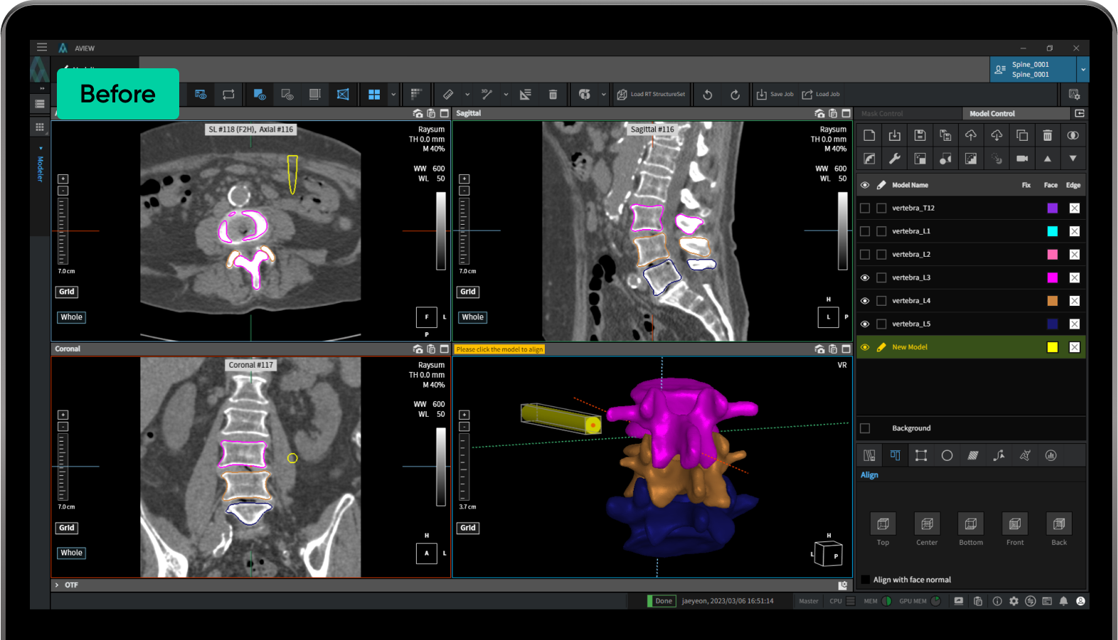
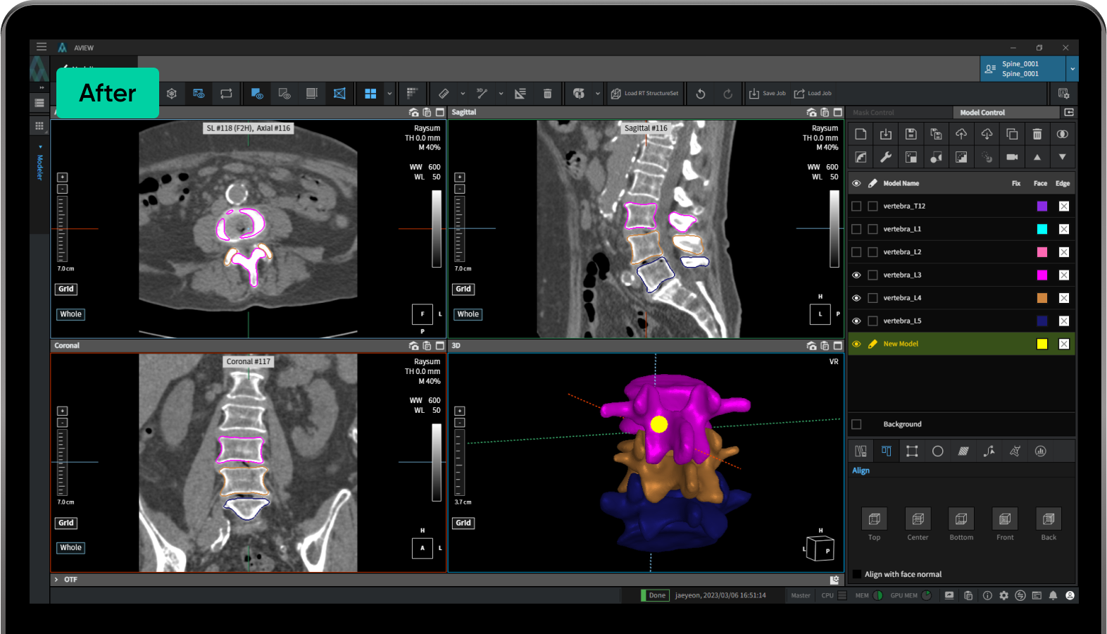
Align Model
Align objects to a desired position.
Transform Model
Move, rotate and adjust size to desired values
02

03
Generate database by purpose
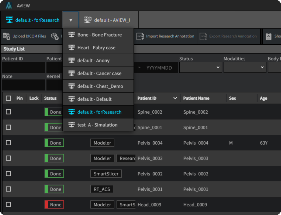
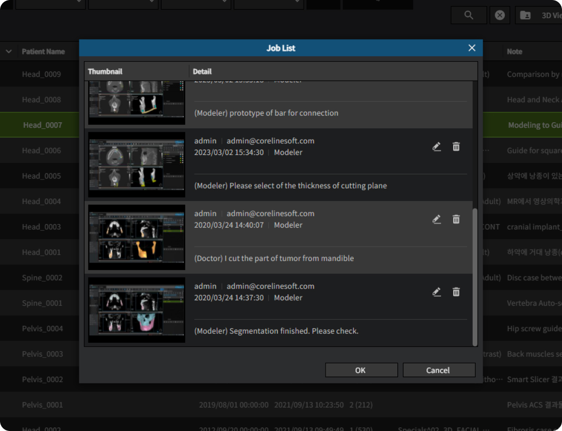
Review Work history by time
04
01
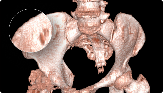
Smart slicerBefore
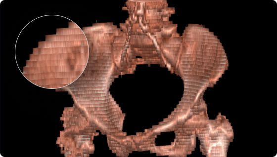
After
02
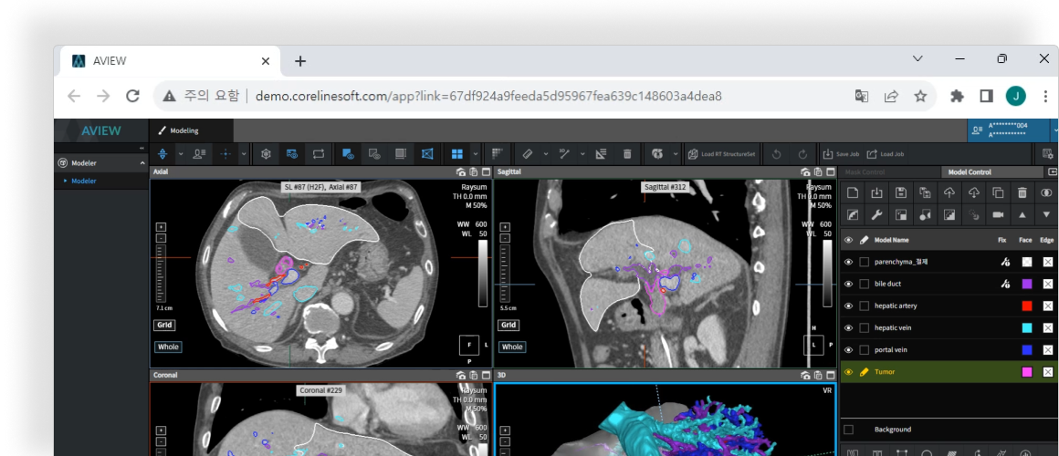
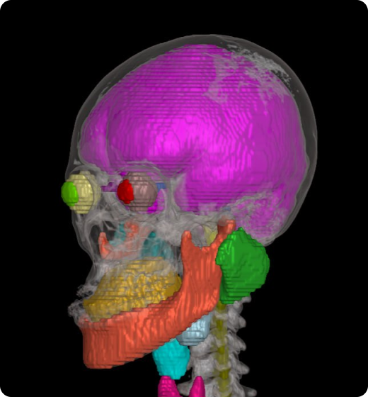
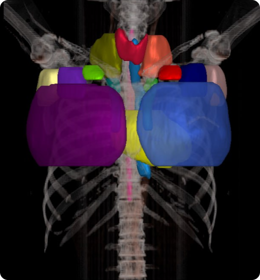
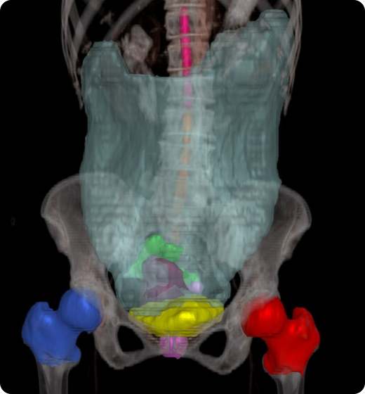
Organ segmentation and contouring functionsHead & Neck
22 Organs
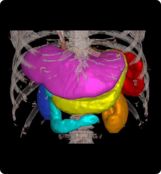
Abdomen
5 Organs
Breast
12 Organs
Pelvis
10 Organs
Publications
Refer to the following research papers
These contents represent summaries of scientific
publications and are unrelated to any form of advertising.

This study aimed at evaluating the difference between amorphous bone fracture (AFF) patients and general population between patients and general population between the general population between patients and general population.
In order to obtain a wide range of 46 patients including geometric analysis software analysis software analysis software, including geometric analysis software analysis software analysis software analysis software, including geometric analysis software.
Importantly CT image using aview Model for the core line software image, and created 3D Sampling. This study found that it has increased from executives AFF, but it found that it is similar to electronic HaFF, but compared to general population.
Jung IJ, Kim JW. “Differences in femur geometry and bone markers in atypical femur fractures and the general population.” Sci Rep. 2021 Dec 17;11(1):24149.

The goal of this study is to develop an automated method of imaging and tracking the location of the Hachijo nerve (IAN) using artificial intelligence (AI) on the cone beam computed tomography data set.
We used customized 3D nnU-Net for image segmentation, and we repeatedly performed active learning on 83 datasets. Using the remaining 50 datasets, we evaluated the accuracy of the model for IAN segmentation and compared the Dice Similarity Factor (DSC) values and segmentation times for each stage of learning.
Deep active learning frameworks have been found to be fast, accurate and powerful clinical tools for distinguishing IAN locations.
The full segmentation was performed using the aview Modeler, and ground truth in IAN was provided by three experts.
Lim HK, Jung SK, Kim SH, Cho Y, Song IS. “Deep semi-supervised learning for automatic segmentation of inferior alveolar nerve using a convolutional neural network.” BMC Oral Health. 2021 Dec 7;21(1):630
Intended purpose
Intended user
Warning
Caution