

01
02


03
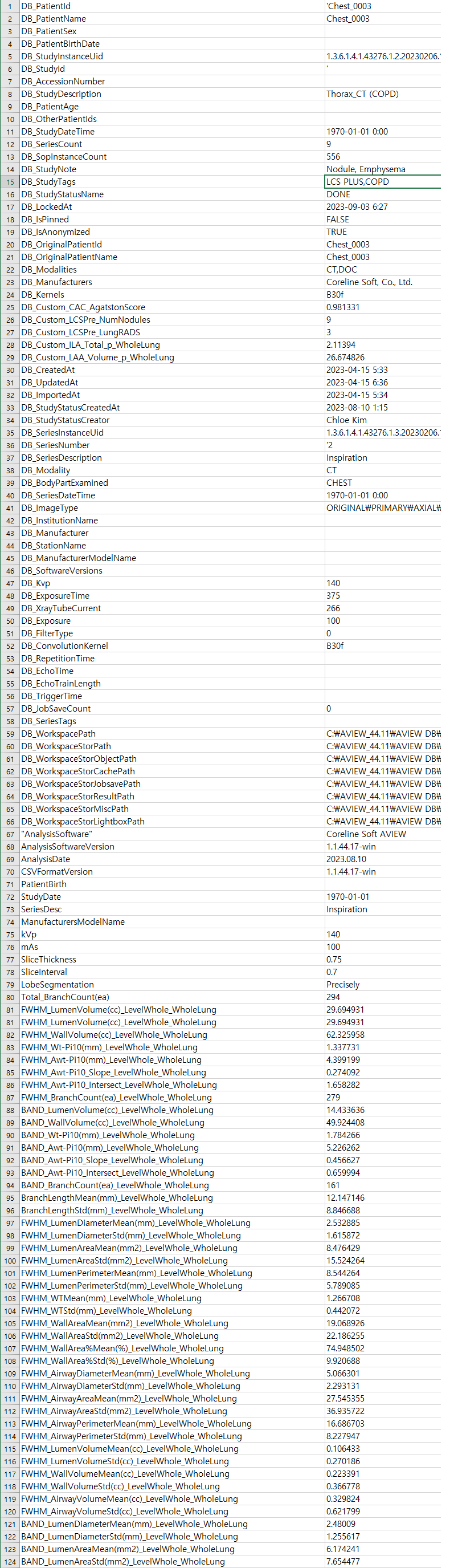
Analysis Results in CSV

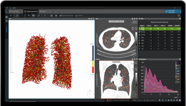
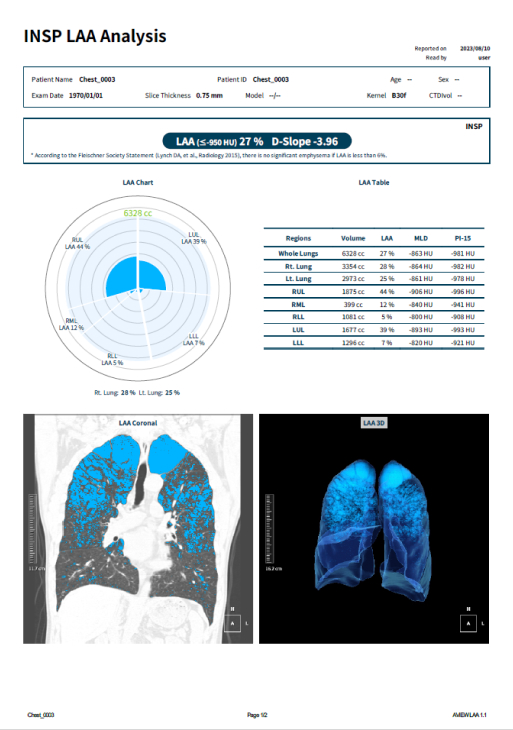
Report
Publications
Refer to the following research papers
These contents represent summaries of scientific
publications and are unrelated to any form of advertising.

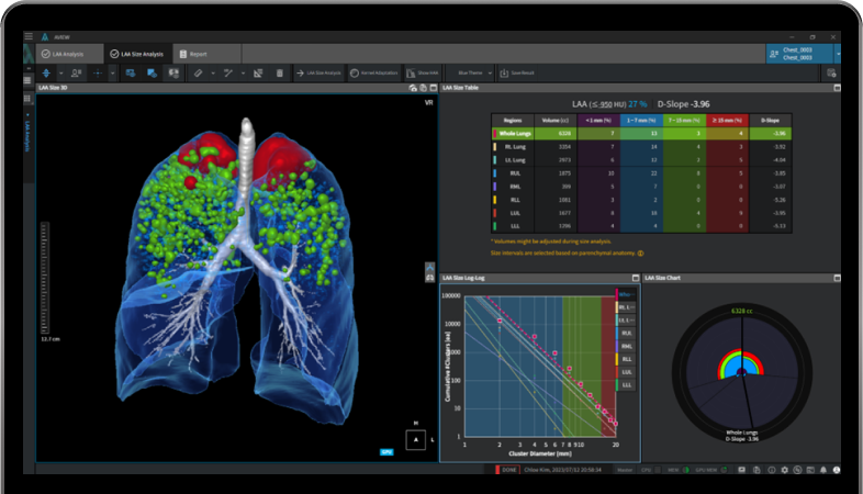
Quantitative CT EAtC mapping was performed using an automatic segmentation software (aview, Coreline Soft). Lung segmentation was performed for EAtC mapping, and the airways, vessels, and background were segmented and removed from the lung parenchyma using several specific thresholds. The proposed quantitative CT EAtC mapping provides comprehensive lung functional information on each disease component of COPD, which may serve as an imaging biomarker of lung function.
Hwang HJ, Seo JB, Lee SM, Kim N, Yi J, Lee JS, Lee SW, Oh YM, Lee SD. New Method for Combined Quantitative Assessment of Air-Trapping and Emphysema on Chest Computed Tomography in Chronic Obstructive Pulmonary Disease: Comparison with Parametric Response Mapping. Korean J Radiol. 2021 Oct;22(10):1719-1729. doi: 10.3348/kjr.2021.0033. Epub 2021 Jul 1. PMID: 34269529; PMCID: PMC8484152.

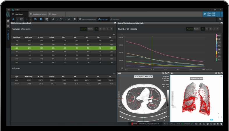
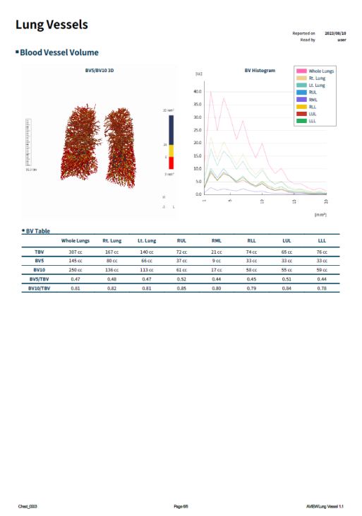
A total of 30 patients with CTEPH who received multimodal treatment, including riociguat and underwent both non-contrast CT for pulmonary vasculature analysis and RHC pre- and post-treatment were included Lung segmentation and pulmonary vasculature analyses were performed using the aview® system (Coreline Soft) Non-contrast CT measures could quantitatively assess changes in the pulmonary vasculature in response to treatment and were correlated with hemodynamic and clinical parameters.
Huang YS, Chen ZW, Lee WJ, Wu CK, Kuo PH, Hsu HH, Tang SY, Tsai CH, Su MY, Ko CL, Hwang JJ, Lin YH, Chang YC. Treatment Response Evaluation by Computed Tomography Pulmonary Vasculature Analysis in Patients With Chronic Thromboembolic Pulmonary Hypertension. Korean J Radiol. 2023 Apr;24(4):349-361. doi: 10.3348/kjr.2022.0675. Epub 2023 Mar 7. PMID: 36907594; PMCID: PMC10067691.
Intended purpose
Intended user
Warning
Caution