

Conformity Certifications
MFDS
Republic of Korea
TGA
Australia
HSA
Singapore
ANVISA
Brazil
HC
Canada
CE
Europe
01
02
Report
Validations
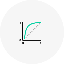
We have successfully demonstrated the identification of interstitial lung abnormalities
In advanced research findings, when employing an ILA threshold of 1.8%...

0
Sensitivity

0
Specificity
0
AUROC
Chae KJ, Lim S, Seo JB, Hwang HJ, Choi H, Lynch D, Jin GY. Interstitial Lung Abnormalities at CT in the Korean National Lung Cancer Screening Program: Prevalence and Deep Learning-based Texture Analysis. Radiology. 2023 May;307(4):e222828.
Publications
Refer to the following research papers
These contents represent summaries of scientific
publications and are unrelated to any form of advertising.

The object of this study was to determine the prevalence of ILA at CT examinations from the Korean National Lung Cancer Screening Program (participants who underwent chest CT between April 2017 and December 2020) and define an optimal lung area threshold for ILA detection with CT with use of deep learning–based texture analysis.
ILA were detected in 4% of the Korean lung cancer screening population. Deep learning–based texture analysis (Aview ILA, Corelinesoft) showed high sensitivity and specificity for detecting ILA with use of a 1.8% lung area cutoff value.
Chae KJ, Lim S, Seo JB, Hwang HJ, Choi H, Lynch D, Jin GY. Interstitial Lung Abnormalities at CT in the Korean National Lung Cancer Screening Program: Prevalence and Deep Learning-based Texture Analysis. Radiology. 2023 May;307(4):e222828.

The object of this study was to evaluate the performance of a fully automated quantitative software (Aview ILA, Corelinesoft) in detecting interstitial lung abnormalities (ILA) according to the Fleischner Society guidelines on routine chest CT compared with radiologists' visual analysis.
The quantification system for identifying ILA using a threshold of 5 % in at least one zone showed 67.6 % sensitivity, 93.3 % specificity, and 90.5 % accuracy.
The best cut-off value of abnormality extent detecting ILA on the system was 3.6 %. Inter-reader agreement was substantial for ILA but only fair for its subtypes. Applying an automated quantification system in routine clinical practice may aid the objective identification of ILA.
Kim MS, Choe J, Hwang HJ, Lee SM, Yun J, Kim N, Ko MS, Yi J, Yu D, Seo JB. Interstitial lung abnormalities (ILA) on routine chest CT: Comparison of radiologists' visual evaluation and automated quantification. Eur J Radiol. 2022 Dec;157:110564.