When undergoing a chest CT scan for lung cancer screening, many people become concerned upon hearing the term 'lung nodule.' However, a lung nodule does not necessarily mean lung cancer. This article aims to clarify misconceptions and provide accurate information by explaining the types and causes of lung nodules and their relationship to lung cancer.
What is a Lung Nodule?
A lung nodule primarily refers to an abnormal mass observed within the lung that is 3cm or less in size. If the lesion exceeds 3cm, it is referred to as a tumor or mass rather than a nodule. It is important to note that a lung nodule itself is not a disease name but rather an imaging finding observed on a chest CT or X-ray due to various underlying conditions. Most lung nodules do not cause any specific symptoms and are often discovered incidentally during health check-ups via chest CT scans.
Lung Nodule: Benign or Malignant?
The main causes of lung nodules are highly diverse and can be broadly categorized into benign conditions, such as infections, and malignant conditions, such as cancer. Benign causes can include pneumonia, tuberculosis, fungal infections, and benign tumors (hamartomas), while lung cancer or metastatic cancer are classified as malignant nodules. It is often challenging to distinguish between these various causes based on imaging alone. In Korea, particular caution is needed during differential diagnosis, as tuberculomas caused by pulmonary tuberculosis are common.
While lung nodules themselves are unlikely to cause specific complications, if a nodule continuously grows, it can compress the bronchi and lead to complications such as pneumonia. More importantly, a lung nodule could be an early sign of lung cancer. Therefore, even small nodules require regular follow-up, and if a nodule is large or highly suspected of being malignant, a biopsy should be performed for an accurate diagnosis.
The frequent incidental discovery of small lung nodules on health check-up CT scans, coupled with the uncertainty of whether they are benign or malignant, can cause significant anxiety and psychological burden for those screened. Therefore, accurate interpretation by a radiologist, along with clear and empathetic counseling from medical staff, is crucial. This demonstrates that the screening process goes beyond merely diagnosing a disease; it also plays a vital role in ensuring the patient's psychological well-being and reducing unnecessary fear.
What is Ground-Glass Opacity (GGO)?
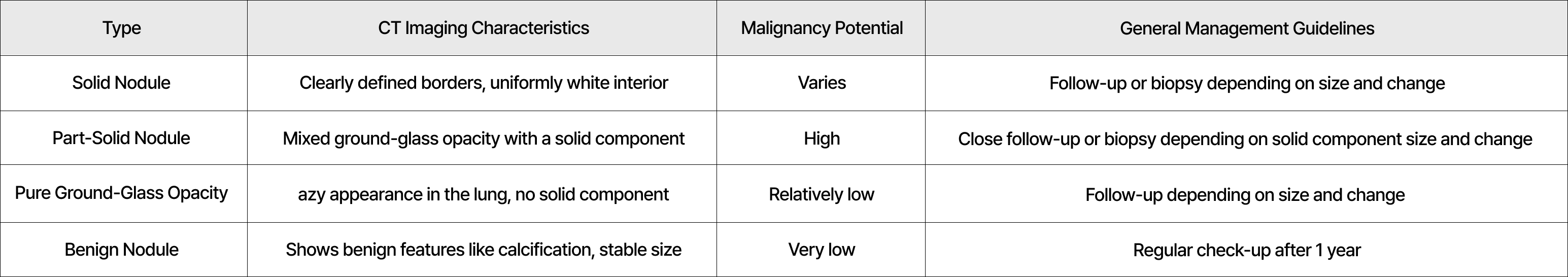
Ground-glass opacity (GGO) is an imaging term referring to a hazy, cloudy area in the lung on a chest CT scan. It is not a specific disease name and is broadly classified into two types based on the presence of solidification: 'pure ground-glass opacity' (no solidification) and 'mixed ground-glass opacity' (part-solid nodule) with some solidification.
Ground-glass opacity can be an early form of lung cancer, requiring particularly careful follow-up. The likelihood of malignancy increases, especially if a solid component newly appears or grows, if the size of the ground-glass opacity increases, or if, in the case of mixed ground-glass opacity, the solid component is 8mm or larger on the initial scan, or increases by 4mm or more on follow-up scans.
Lung nodules vary in their malignant potential depending on their size and characteristics (solid, part-solid, ground-glass opacity), and follow-up guidelines are accordingly subdivided. This precise classification and management guideline demonstrate that lung cancer screening is not just about simple screening but implements the concept of 'precision medicine' from the initial diagnostic stage. This emphasizes the importance of a personalized approach optimized for each patient's risk.
Other Significant Findings Beyond Lung Nodules
Chest CT scans can reveal various other chest abnormalities besides lung nodules. Even if not directly related to lung cancer, other lung diseases or chest conditions requiring further examination or treatment may be found. Chest CT plays a crucial role in identifying the presence, size, and metastasis of a wide range of lung and chest diseases, including pulmonary tuberculosis, pneumonia, emphysema, bronchiectasis, pleurisy, mediastinal diseases, and lymph node lesions, in addition to lung cancer. Therefore, a precise understanding of the CT scan results and consultation with medical professionals are essential.
Types and Characteristics of Lung Nodules
The interpretation and management of lung nodules are more critical than their mere discovery. While most are benign, some can be an early form of lung cancer, necessitating accurate information and planned follow-up rather than excessive anxiety.
The key is accurate imaging interpretation combined with a tailored response to any changes observed. Furthermore, if significant abnormalities are found, it is essential to discuss the next steps with a medical professional for an accurate diagnosis. This marks the starting point for precise and systematic patient-centered management of lung diseases, extending beyond simple CT interpretation.
Lung Cancer Screening Series
1. The Importance of Lung Cancer Screening: Early Detection Changes Survival Rates
2. Mastering Lung Cancer Screening Methods: What is Low-Dose CT (LDCT)?
3. (This article) Lung Nodules Found on Lung Scans – Are They Always Lung Cancer?
4. Lung Cancer Screening Eligibility Checklist: Do You Qualify?
5. Interpreting Lung CT Scan Results: Understanding Lung-RADS
6. Common Questions About Lung Cancer Screening: Myths and Facts
7. The Future of Lung Cancer Screening: How AI and Liquid Biopsy are Changing the Landscape
8. National Lung Cancer Screening: Do I Need It? Age and Cycle Summary


 List
List